
Many patients receive a Doppler scan referral without a clear understanding of what the test actually does or why it was recommended over other imaging options. Unlike a standard ultrasound that shows anatomical structure, a Doppler scan goes further — it shows how blood is moving through your vessels in real time. That distinction matters enormously for the clinical conditions it is used to evaluate.
If your doctor has recommended a Doppler scan in Bangalore, understanding what the test involves, how to prepare, and what to expect from a credible diagnostic centre will help you approach the appointment with clarity rather than anxiety.
How Doppler Ultrasound Works
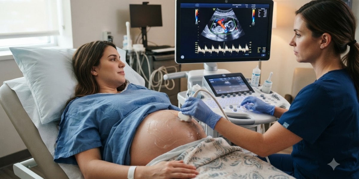
A Doppler scan uses the Doppler effect — the same principle behind a car horn sounding different as it approaches and moves away — applied to sound waves and blood flow. The ultrasound machine detects the frequency shift of sound waves bouncing off moving red blood cells. This creates a visual representation of blood flow direction, speed, and turbulence.
There are three main types used clinically:
Colour Doppler — Displays blood flow in colour overlay on a standard ultrasound image; arteries and veins shown in different colours
Power Doppler — More sensitive to low blood flow, used for detecting flow in small vessels
Spectral (Pulsed Wave) Doppler — Produces a waveform graph showing flow velocity over time, used for precise haemodynamic assessment
The type your doctor orders depends on what they are trying to assess.
Why Doppler Imaging Is Clinically Important
Conditions Commonly Investigated with Doppler
A Doppler scan in Bangalore is ordered across multiple clinical scenarios. The most common include:
Vascular conditions:
Deep vein thrombosis (DVT) — detecting blood clots in leg veins
Peripheral arterial disease — assessing narrowing of arteries in the limbs
Carotid artery stenosis — evaluating blockage risk for stroke
Varicose vein assessment — identifying faulty valves in superficial veins
Obstetric and gynaecological conditions:
Foetal wellbeing — monitoring blood flow through the umbilical cord and foetal brain
Placental insufficiency — evaluating nutrient and oxygen delivery to the foetus
Ovarian torsion — detecting blood flow changes in the ovary
Abdominal conditions:
Portal hypertension — liver disease assessment
Renal artery stenosis — evaluating kidney blood supply
Post-transplant evaluation — checking vessel patency in transplanted organs
Cardiac adjacent applications:
Echocardiography often uses Doppler principles to assess heart valve function and blood flow across chambers
The wide application of Doppler imaging makes it one of the most versatile diagnostic tools available.
What Happens During a Doppler Scan
The procedure is non-invasive, painless, and does not use radiation. Here is what typically happens:
You will lie on an examination table and the sonographer will apply a water-based gel to the skin over the area being examined
The transducer (a handheld probe) is pressed gently against the skin and moved over the area
You will hear sounds — these are the actual blood flow sounds converted to audio feedback that the sonographer uses alongside the visual display
The sonographer captures images and flow measurements across different points
The session typically lasts between 20 and 45 minutes depending on the complexity and the body region being examined
No needles, no radiation, no special recovery period.
Preparation Requirements by Scan Type
Preparation for a Doppler scan varies based on the region being examined:
Scan Type | Preparation Required |
Carotid Doppler | No preparation needed |
Peripheral Venous/Arterial | Avoid compression stockings; wear loose clothing |
Abdominal/Portal Doppler | Fasting for 4–6 hours (reduces bowel gas) |
Renal Doppler | Fasting for 4–6 hours; drink water before to fill the bladder |
Obstetric Doppler | Usually no fasting; follow your doctor's instructions |
Your booking confirmation from the diagnostic centre should specify the exact preparation for your scan. If it does not, call and ask before your appointment.
Choosing a Doppler Scan Centre in Bangalore
Not all ultrasound machines have equal Doppler capability. Older machines may have limited colour Doppler sensitivity, particularly for evaluating low-flow states or small vessels. This is relevant for detecting early DVT or assessing foetal vessels in pregnancy.
When evaluating Doppler scan services in Bangalore, consider the following:
Sonographer experience: Doppler interpretation is highly operator-dependent. A skilled sonographer knows how to optimise probe angle, scan depth, and colour gain to capture accurate flow data. Experience in vascular or obstetric Doppler specifically matters.
Radiologist reporting: The sonographer performs the scan, but a qualified radiologist or sonologist interprets the findings and writes the report. Confirm that the centre has this process in place rather than having the sonographer self-report.
Equipment modernity: Ask whether the machine supports colour Doppler, power Doppler, and spectral Doppler. A centre that can only offer basic colour Doppler may not capture all the information your doctor needs.
Turnaround time: For conditions such as DVT or foetal distress, same-day reporting is clinically important. Confirm this at booking.
Understanding Your Doppler Report
Doppler reports use terminology that can seem technical. Here are common terms and what they mean:
Peak Systolic Velocity (PSV) — Maximum speed of blood during the heart's pumping phase; elevated in artery narrowing
End Diastolic Velocity (EDV) — Flow speed between heartbeats; absent or reversed flow can indicate serious circulatory problems
Resistive Index (RI) — A ratio measuring vessel resistance; elevated in certain kidney and obstetric conditions
Pulsatility Index (PI) — Another flow resistance measure, commonly used in obstetric Doppler to assess foetal circulation
IMT (Intima-Media Thickness) — Thickness of the carotid artery wall; thickening suggests early atherosclerosis
Your treating doctor will contextualise these values against your clinical history and symptoms.
Frequently Asked Questions
Is a Doppler scan the same as a regular ultrasound?
A regular ultrasound shows anatomical structures — organ size, shape, and texture. A Doppler scan adds real-time blood flow evaluation to this. Many scans combine both in a single appointment, but they serve different diagnostic purposes.
Can a Doppler scan detect a blood clot?
Yes. Venous Doppler is the primary non-invasive test for diagnosing deep vein thrombosis. A clot will show as absent or abnormal blood flow in the affected vessel, and the vein will not compress normally under probe pressure.
Is Doppler scanning safe during pregnancy?
Doppler ultrasound has an extensive safety record in obstetric care. It does not use radiation and the sound wave intensities used are well within established safety guidelines. It is routinely used to monitor foetal wellbeing in high-risk pregnancies.
Do I need a referral for a Doppler scan in Bangalore?
Most diagnostic centres will request a doctor's referral to ensure the appropriate scan type and region are assessed. This also helps the radiologist correlate findings with your clinical history. Self-referral may be possible in some centres but is generally not recommended for vascular assessments.
How quickly will I get my Doppler scan results?
Many centres in Bangalore offer same-day or next-day reports. For urgent clinical situations — particularly obstetric Doppler or suspected DVT — confirm at booking that same-day reporting is available.
A Doppler scan provides information that structural imaging simply cannot — how blood is moving, where it is restricted, and how foetal or vascular health is changing over time. For the conditions it is designed to assess, it remains one of the most clinically direct diagnostic tools available. Choosing a centre with proper equipment and experienced reporting ensures that the information your doctor receives is accurate and actionable.