
The first time you get handed a protein structure assignment, it can feel like you're being asked to learn five subjects at once sequence analysis, structural biology, database navigation, molecular visualization software, and scientific writing, all due in two weeks.
Here's the thing that actually helps: you don't tackle all of it simultaneously. There's a logical order to this work. You identify the protein, you look at its sequence, you find or predict its structure, you connect that structure to what the protein actually does, and then you're honest about how confident you are in each piece of evidence.
Below is the sequence I'd walk a student through, using a real protein hen egg-white lysozyme as a running example so the steps aren't just theoretical.
What a Protein Structure Study Actually Involves
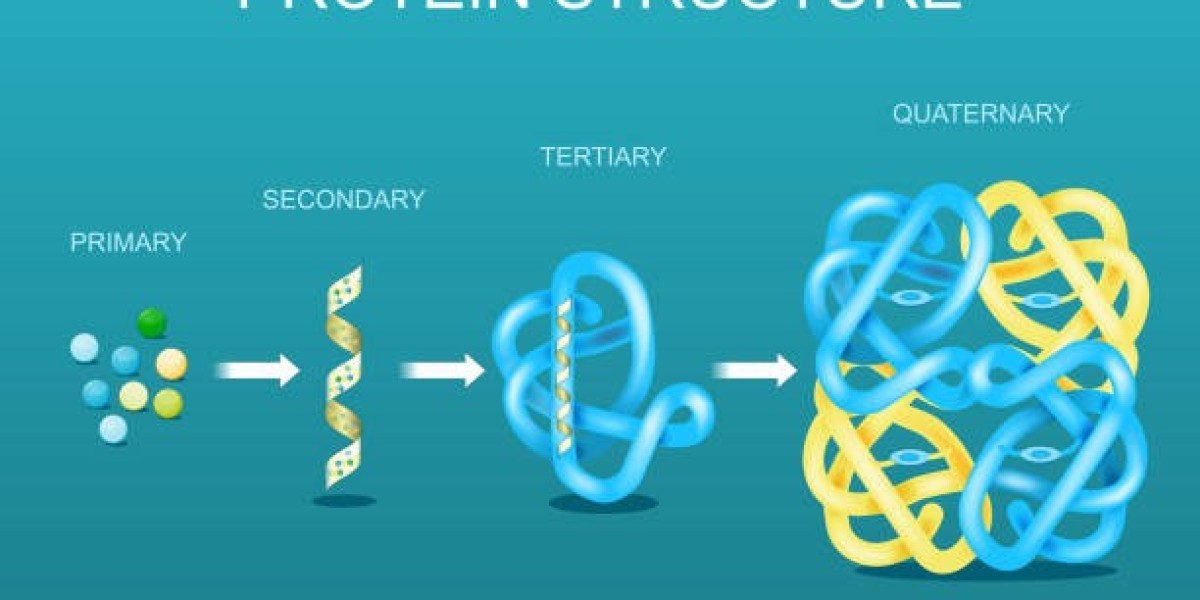
Proteins fold into three-dimensional shapes, and that shape is basically the whole story of what the protein does. Structural biologists usually break this down into four levels:
- Primary structure the amino acid sequence itself
- Secondary structure local folding patterns, mainly alpha-helices and beta-sheets
- Tertiary structure the full 3D shape of a single chain
- Quaternary structure how multiple chains fit together, if the protein has more than one
You probably won't be asked to cover all four in depth. Maybe you're identifying an unknown protein from a sequence. Maybe you're comparing two related enzymes. Maybe you're looking at how a single mutation might mess up a binding site. Whatever the specific task, the goal is the same: don't just describe the protein explain what the evidence tells you about it.
Step 1: Pin Down Exactly Which Protein You're Working With
Before anything else, confirm the protein's identity properly. This sounds obvious, but it's the step people rush through, and it causes problems later.
UniProt is where I'd start. Search for "lysozyme C," then filter by organism Gallus gallus (chicken) for egg-white lysozyme, the classic experimental protein and confirm the entry is reviewed (Swiss-Prot, not TrEMBL). Reviewed entries have been checked by curators against published literature; TrEMBL entries are computationally generated and haven't had that manual pass. That distinction matters more than people realize an unreviewed annotation might be a reasonable guess, not a confirmed fact.
While you're on the entry, write down:
- Protein name and gene name
- Organism
- UniProt accession number
- Sequence length
- Known biological function
- Key residues or active-site information
- Linked experimental structures
- Cited literature
This becomes your reference sheet for everything that follows.
Step 2: Read the Sequence for Clues
The raw amino acid sequence tells you more than it looks like it should.
Stretches of hydrophobic residues often flag membrane-spanning regions. Clusters of conserved residues ones that show up in the same position across related proteins from different species usually matter for binding or catalysis. In lysozyme's case, two residues (a glutamate and an aspartate, positioned within the active-site cleft) are the catalytic pair that actually breaks down bacterial cell walls. That's not something you'd guess from the sequence alone, which is exactly why the next tools matter.
NCBI's Conserved Domain Database will scan the sequence against curated domain models and flag matches. InterPro does something similar but pulls from several signature databases at once, which is useful when one database's model doesn't quite match your protein.
Why bother identifying domains separately?
Because most proteins aren't one uniform blob they're built from distinct functional units. If InterPro tells you there's a catalytic domain and a separate substrate-binding region, you now have something specific to look for once you get to the 3D structure. "This protein has a 3D structure" is not an argument. "This protein's catalytic domain sits adjacent to its binding domain, consistent with X" is.
Step 3: Find (or Locate) an Experimental Structure
Now you go looking for the actual 3D shape.
The Protein Data Bank is where experimentally solved structures live you can search by protein name, accession, or sequence. For lysozyme, you'll get a long list of entries (6LYZ, 1LYZ, 2LYZ, and dozens more), because it's one of the most-studied proteins in structural biology history.
Don't just grab the first result. Check:
- What experimental method was used
- Resolution (if X-ray)
- How many chains are in the structure
- Whether a ligand or substrate is bound
- Any missing residues in the model
- Whether the sequence has engineered mutations
- What the actual biological assembly looks like
- The paper the structure came from
This matters practically. One lysozyme structure might just be the apo form (no ligand bound); another might have a substrate analog sitting in the active site, which is far more useful if you're trying to explain how the enzyme works rather than just what it looks like.
If you need additional academic guidance while working through a project, you may also encounter resources offering bioinformatics assignment writing services uk. If you use external academic support, make sure the resulting work complies with your university's policies on originality, attribution and permitted assistance.
Step 4: Understand How the Structure Was Actually Solved
Don't just name-drop the method in your report explain why it's relevant to how you interpret the model.
- X-ray crystallography gives you a static, high-resolution snapshot, but flexible loops sometimes don't show up clearly (or at all) because they don't hold still enough to diffract cleanly.
- NMR spectroscopy works in solution, so it's better suited to small, flexible proteins, and often gives you an ensemble of slightly different conformations rather than one fixed structure.
- Cryo-EM has gotten dramatically better in recent years and is now the go-to method for large complexes that don't crystallize well.
For a small, well-behaved protein like lysozyme, X-ray crystallography is why we've had reliable structures since the 1960s it was actually the first enzyme structure ever solved this way. That historical detail alone is worth a sentence in an introduction if you want your assignment to read like it was written by someone who understands the field, not just someone who searched a database.
Step 5: When There's No Experimental Structure, Use a Predicted One
Plenty of proteins especially ones outside well-studied model organisms don't have a solved structure yet. That's where AlphaFold comes in.
AlphaFold's prediction accuracy, demonstrated at the CASP14 assessment and published in Nature, was genuinely a turning point for the field. But and this is where a lot of assignments lose marks a predicted structure is not the same category of evidence as an experimentally solved one.
Every AlphaFold model comes with a per-residue confidence score (pLDDT). High-confidence regions are usually reliable; low-confidence regions often flexible loops or disordered segments should be treated with real skepticism. Use the prediction as a hypothesis-generating tool, not as proof of anything.
Step 6: Actually Look at the Structure
A static image of a protein tells you almost nothing on its own. You need to move around it.
UCSF ChimeraX is the standard free tool for this. Load your PDB file, and you can switch between representations depending on what you're trying to show:
- Cartoon/ribbon view best for seeing secondary structure (you'll spot lysozyme's mix of alpha-helices and a small beta-sheet region immediately)
- Surface view shows overall shape and which regions are exposed versus buried
- Stick representation for zooming into individual residues, like the catalytic glutamate-aspartate pair in the active site
Don't screenshot a pretty ribbon diagram just because it looks scientific. Every figure you include should be doing a specific job pointing at something the reader wouldn't otherwise notice.
Step 7: Connect the Structure Back to Function
This is the part that separates a descriptive report from an actual analysis.
For lysozyme, you'd want to show the active-site cleft, point out the two catalytic residues, and explain how their spatial arrangement lets the enzyme hydrolyze the glycosidic bonds in bacterial peptidoglycan. If your assignment involves a membrane protein instead, you'd be looking at transmembrane segments and how intracellular versus extracellular domains are arranged. For a DNA-binding protein, you'd focus on the structural motifs that make contact with the nucleic acid backbone.
Whatever the protein, use UniProt and InterPro to find out which regions already have experimental support behind their functional annotation before you go hunting for them in the 3D model. That way you're confirming known biology with structural evidence, not just speculating.
Step 8: Compare It Against a Related Protein
If your assignment allows for it, pulling in a second, related protein adds real depth. Compare lysozyme C against a related lysozyme isoform, or against a homolog from a different organism, and ask:
- Are the major domains arranged the same way?
- Are the catalytically important residues conserved?
- Does the active site have a similar shape and chemistry?
- Are there extra domains in one protein but not the other?
- Could any structural differences explain a functional difference?
"These two proteins look similar" isn't an argument. "These two proteins share a conserved catalytic dyad but differ in loop length near the substrate-binding groove, which may affect specificity" is.
Step 9: Be Honest About the Quality of Your Evidence
Not every piece of information you gather carries the same weight.
An experimentally solved structure is direct evidence. A curated UniProt annotation is usually a summary of published experimental work. A computational prediction is a hypothesis, however good the underlying model is.
For experimental structures, check the validation reports available through PDB and PDBe they'll flag unusual geometry or low-confidence regions. For AlphaFold models, actually look at the confidence scores instead of ignoring them. A report that quietly glosses over these limitations reads as less credible, not more markers notice when uncertainty gets hidden.
A Reasonable Structure for the Written Report
- Introduction the protein, and why its structure matters
- Sequence analysis accession, length, notable sequence features
- Domain and functional analysis conserved domains and known functional regions
- Structural analysis the experimental structure or predicted model, and its key features
- Structure–function relationship the actual analytical core of the assignment
- Comparison a related protein or homolog, if the brief allows it
- Limitations missing data, prediction confidence, experimental caveats
- Conclusion what you actually found, without overreaching
Common Ways This Kind of Assignment Loses Marks
- Copy-pasting database descriptions. Interpreting is the assignment; summarizing someone else's summary isn't.
- Mixing up prediction and experiment. Always state explicitly which one you're looking at.
- Skipping the limitations section. Every model has gaps pointing them out shows you understand the data, not just the software.
- Including images with no explanation. Every figure needs a caption that tells the reader what to look for.
- Overstating conclusions. If the structure suggests a mechanism but nothing confirms it experimentally, say "suggests" or "may indicate" not "shows."
Quick Checklist Before You Submit
- Have you confirmed the correct protein and a reliable sequence source?
- Have you identified its domains and key residues?
- Do you know where your structural model actually came from?
- Have you clearly separated experimental data from prediction?
- Have you explained what the structure shows, not just displayed it?
- Have you tied structural features back to biological function?
- Have you acknowledged the limitations of your evidence?
- Are your figures captioned and purposeful?
The Bottom Line
A solid protein structure assignment is a small research investigation, not a database scavenger hunt. You start with a sequence, use the major databases to understand how the protein is organized, examine a structure solved or predicted and then build an argument about what that structure tells you about function.
The strongest assignments aren't the ones with the most screenshots. They're the ones that make a clear, well-supported argument and are upfront about where the evidence runs out.